
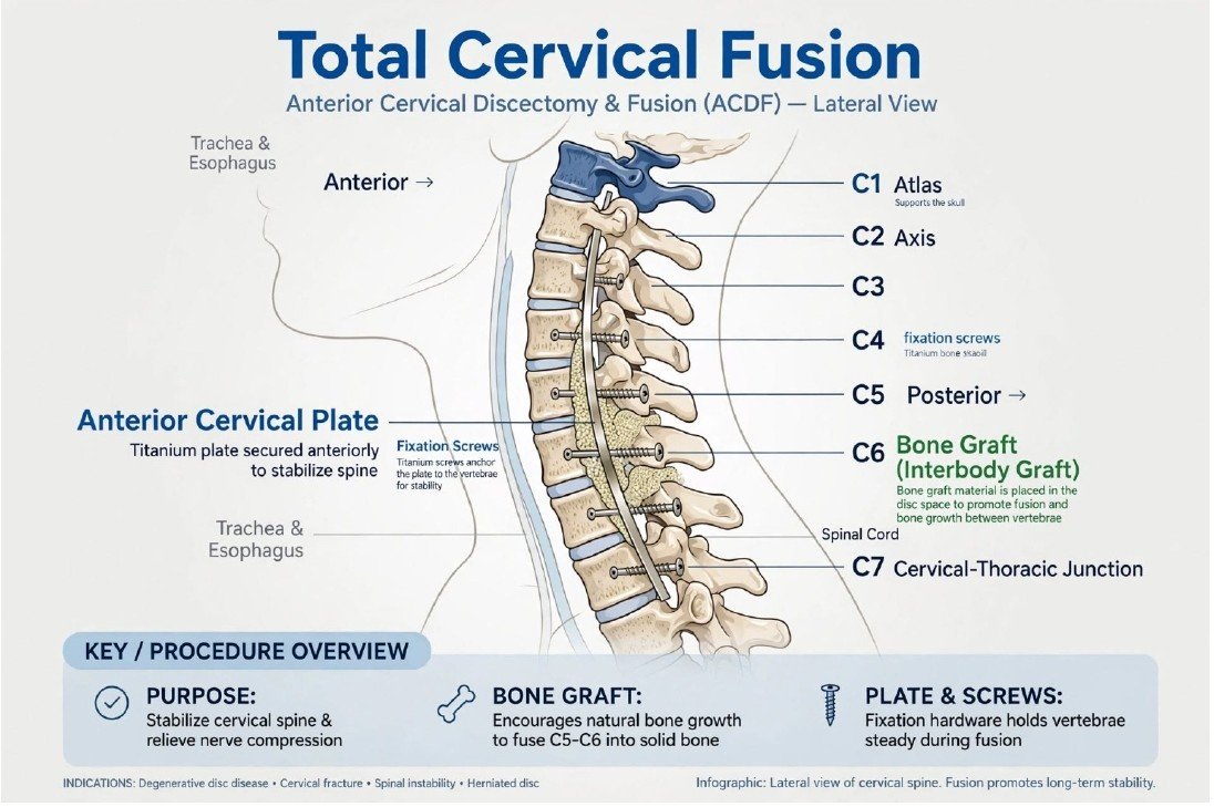
Cervical Discectomy and Fusion (ACDF)
This operation treats selected cases of nerve or spinal cord compression in the neck. Through an incision at the front of the neck, the surgeon removes the affected disc and any bone spurs contributing to compression.
A graft or cage is placed in the disc space to support fusion between adjacent vertebrae. A plate and screws may provide stability while the bones heal together. The aim is to relieve nerve-related symptoms and stabilise the treated levels; recovery and movement restrictions depend on the extent of surgery.
Illustration clarification: The title “Total Cervical Fusion” is misleading: ACDF treats selected levels, not necessarily the whole neck. The drawn plate, graft and vertebral labels are inconsistent with an accurate anterior fusion.
Sources: AAOS: cervical radiculopathy surgical treatment options
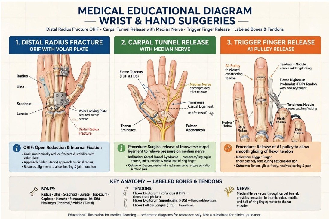
Wrist and Hand Surgery
Distal radius fracture fixation (ORIF): The surgeon realigns a displaced fracture near the wrist and secures the fragments with a plate and screws. A volar plate is placed on the palm-facing surface of the distal radius. Fixation supports healing and recovery of wrist function.
Carpal tunnel release: Dividing the transverse carpal ligament reduces pressure on the median nerve. It may improve numbness, tingling and pain associated with carpal tunnel syndrome. Recovery depends partly on the severity and duration of nerve compression.
Trigger finger release: The tight A1 pulley near the base of the affected finger is released so the flexor tendon can glide more freely. This aims to reduce painful catching or locking.
Illustration clarification: The wrist plate and several bone labels are misplaced. The A1 pulley is near the knuckle at the base of the finger, not near the fingertip as suggested by the drawing.
Sources: AAOS: distal radius fractures broken wrist | AAOS: carpal tunnel syndrome | AAOS: trigger finger
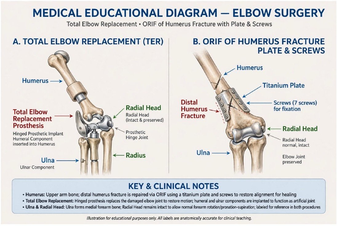
Elbow Replacement and Fracture Fixation
Total elbow replacement: Damaged joint surfaces are replaced with artificial components anchored in the humerus and ulna. It may be considered for severe arthritis or selected complex fractures. The aim is to reduce pain and improve function. Long-term lifting restrictions are commonly required to protect the implant.
Distal humerus fracture fixation (ORIF): Broken fragments at the lower end of the upper arm bone are realigned and held with plates and screws. The aim is to restore joint alignment and allow healing while preserving the natural elbow when possible. Guided rehabilitation helps limit stiffness.
Illustration clarification: The implant shapes, bone labels and fixation positions are schematic and partly inaccurate. Fracture fixation alone does not routinely replace the elbow joint surfaces, although the right panel suggests a metal joint.
Sources: AAOS: total elbow replacement | AAOS: distal humerus fractures of the elbow
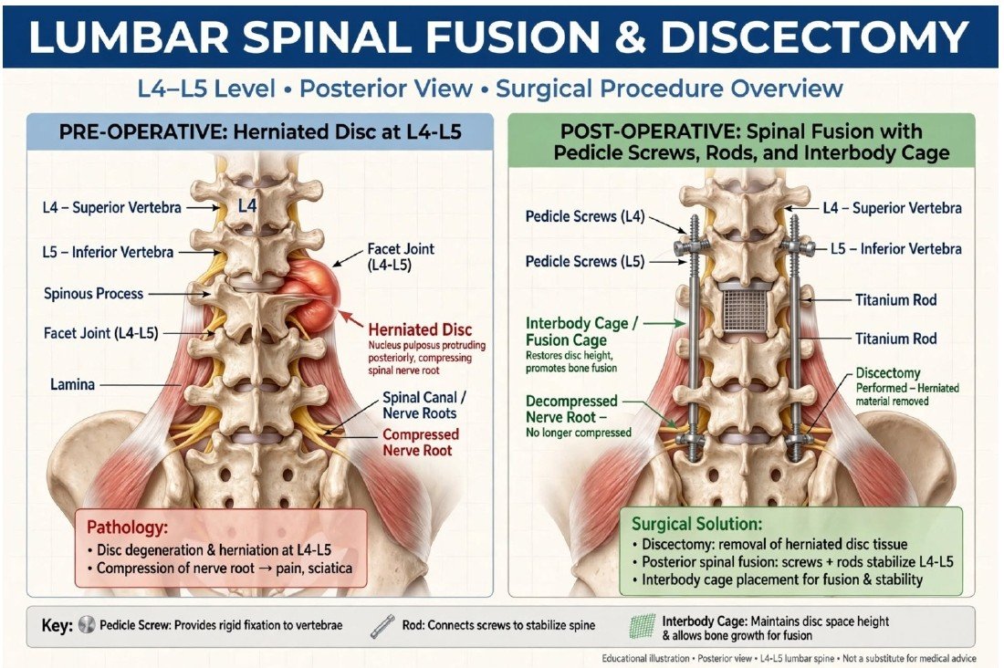
Lumbar Discectomy and Spinal Fusion
Lumbar discectomy: The surgeon removes disc material compressing a nerve in the lower back. This can relieve nerve-related leg pain, such as sciatica, and may help weakness or numbness in appropriately selected patients.
Lumbar fusion: When stabilisation is needed, two or more vertebrae are joined using bone graft. An interbody cage may support the disc space, while screws and rods hold the vertebrae steady duringhealing. Fusion reduces movement at the treated level. A simple disc herniation does not automatically require fusion.
Illustration clarification: The image is labelled L4–L5, but the levels and hardware do not consistently match a single-level fusion. An interbody cage belongs between vertebral bodies in the disc space, not on the back of the spine as drawn.
Sources: AAOS: spinal fusion | AAOS: herniated disk in the lower back
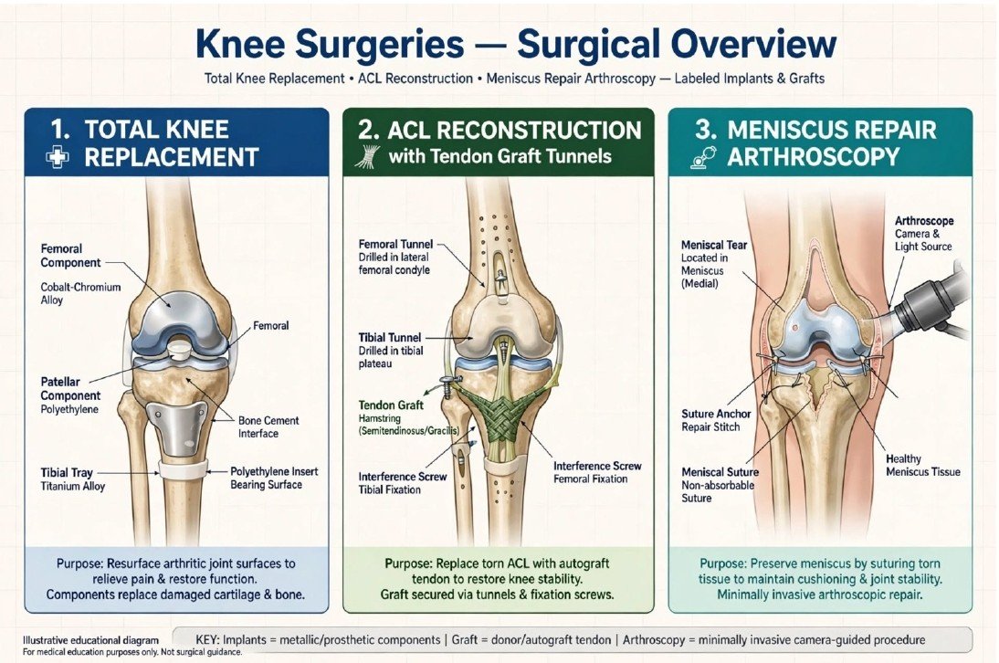
Knee Replacement and Sports Injury Surgery
Total knee replacement: Worn joint surfaces are reshaped and covered with metal components, with a plastic bearing between them. The back of the kneecap may also be resurfaced. The goal is to reduce severe arthritic pain and improve everyday movement.
ACL reconstruction: A torn anterior cruciate ligament is replaced with a tendon graft secured within bone tunnels in the femur and tibia. It aims to improve stability in a knee that gives way. Rehabilitation is essential before returning to demanding activities.
Arthroscopic meniscus repair: Through small incisions, the surgeon uses a camera and instruments to stitch a repairable meniscal tear. This preserves cushioning tissue. Not every tear can be repaired; treatment depends on tear pattern, blood supply and tissue quality.
Illustration clarification: Some replacement components are mislabelled. The ACL graft should run within the joint between the femur and tibia; the external crossing grafts in this diagram are not a correct depiction of standard ACL reconstruction.
Sources: AAOS: total knee replacement | AAOS: acl injury does it require surgery | AAOS: meniscus repair
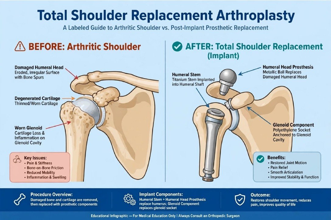
Total Shoulder Replacement
An anatomical total shoulder replacement treats selected patients with painful, severely damaged shoulder joint surfaces. The surgeon replaces the humeral head with an artificial ball and resurfaces the glenoid socket with a prosthetic component.
The operation aims to reduce pain and improve shoulder function. The type of replacement depends on the condition of the joint and rotator cuff. A reverse shoulder replacement may be more suitable in some patients with major rotator cuff damage. Recovery includes protection of healing tissues followed by a staged rehabilitation programme.
Illustration clarification: The humeral head and glenoid labels are misplaced in the “before” panel. The “after” panel shows a detached ball: in an anatomical replacement, the ball connects to the humeral component and sits against the socket.
Sources: AAOS: shoulder joint replacement
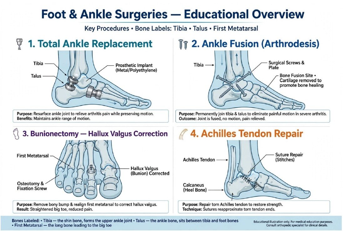
Foot and Ankle Surgery
Total ankle replacement: Damaged ankle joint surfaces are replaced with metal and plastic components. It can reduce arthritic pain while preserving some ankle movement in suitable patients.
Ankle fusion (arthrodesis): The surgeon removes damaged cartilage and stabilises the tibia and talus so they heal together. This aims to reduce painful movement, but movement at the fused ankle joint is lost.
Bunion correction: Surgery realigns the big toe and first metatarsal using bone cuts, soft-tissue correction or selected fusion procedures. Removing the bump alone is often insufficient to correct the deformity.
Achilles tendon repair: Torn tendon ends are stitched together or reattached to the heel bone when appropriate. The goal is to restore the tendon connection used for walking and pushing off. Protected healing and rehabilitation follow surgery.
Illustration clarification: Several implants and labels are misplaced. An ankle replacement sits at the tibia–talus joint; bunion correction concerns the big-toe side; the Achilles tendon attaches behind the heel, not across the top of the foot.
Sources: AAOS: arthritis of the foot and ankle | AAOS: bunion surgery | AAOS: achilles tendon rupture tear
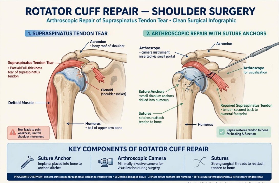
Arthroscopic Rotator Cuff Repair
Rotator cuff repair treats selected tears of the shoulder tendons, including the supraspinatus. Using a small camera and instruments through small incisions, the surgeon assesses the tear and reattaches repairable tendon to its attachment on the upper arm bone using sutures and anchors.
The aim is to reduce pain and improve strength and shoulder function. A sling and staged physiotherapy help protect the repair while the tendon heals. Recovery takes time, and the result depends on factors such as tear size, tissue quality and rehabilitation. Some tears need a different approach or cannot be fully repaired.
Illustration clarification: The tendon path and several labels are inaccurate. The supraspinatus passes beneath the acromion and attaches to the greater tuberosity of the humerus. Anchors secure the tendon at its bony attachment, rather than in the joint’s cartilage surface.
Sources: AAOS: rotator cuff tears surgical treatment options
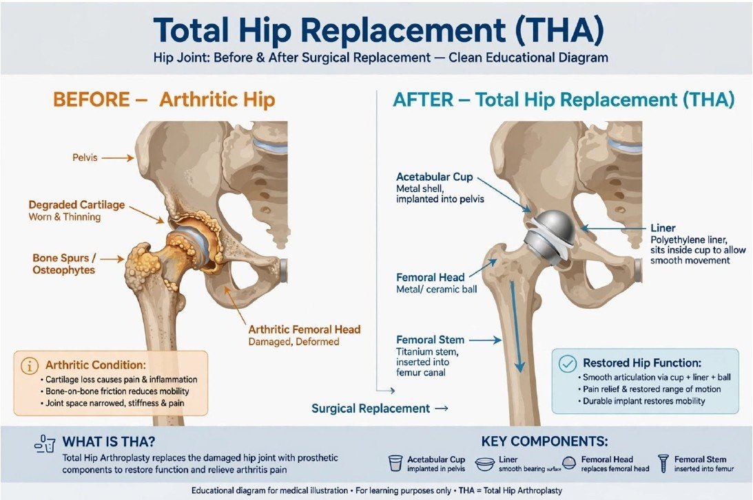
Total Hip Replacement (THA)
Total hip replacement removes the damaged femoral head and replaces it with an artificial ball attached to a stem fitted into the thighbone. The damaged socket is resurfaced with a cup and liner, creating a new bearing surface.
It may be considered when arthritis or other joint damage causes persistent pain and disability despite appropriate nonsurgical care. The aim is to reduce pain and improve walking and daily activities. Rehabilitation, prescribed clot-prevention measures and surgeon-specific precautions support recovery.
Illustration clarification: Some arrows and implant parts are misplaced. The artificial ball connects to the femoral stem and articulates inside the pelvic socket’s liner; the supplied drawing does not accurately show this assembly.
Sources: AAOS: total hip replacement