
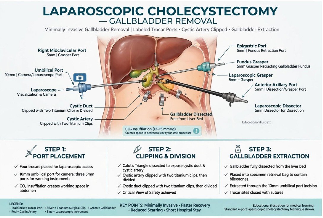
Laparoscopic Gallbladder Removal
Laparoscopic cholecystectomy removes the gallbladder through small abdominal incisions using a camera and surgical instruments. It is commonly performed for symptomatic gallstones or gallbladder inflammation. The surgeon identifies and secures the cystic duct and artery, separates the gallbladder from the liver and removes it.
The liver continues producing bile after surgery, and bile flows directly into the intestine. Some operations need a larger incision for safety. Recovery and dietary advice depend on the patient and any complications.
Illustration clarification: The gallbladder is duplicated and several duct, vessel and port labels are misplaced. Port sizes, clip numbers and extraction sites vary; the diagram is not a precise operative guide.
Sources: Mayo Clinic
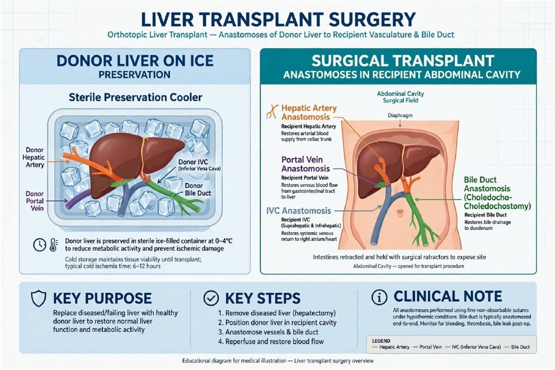
Liver Transplant Surgery
A liver transplant replaces a failing or severely diseased liver with a healthy donor liver, or a portion of one. It may be considered for advanced liver disease, acute liver failure or selected liver cancers after specialist assessment.
The surgeon connects the donor liver to the recipient’s blood vessels and reconstructs bile drainage.Donor tissue can come from a deceased donor or a suitable living donor. Long-term anti-rejectionmedicines and follow-up are required. Availability and suitability depend on transplant evaluation anddonor factors.
Illustration clarification: Vessels and bile ducts are simplified and partly misplaced. A donor liver is protected in sterile preservation packaging or an appropriate perfusion system, not placed directly against loose ice as illustrated. Preservation time and reconstruction methods vary.
Sources: Mayo Clinic
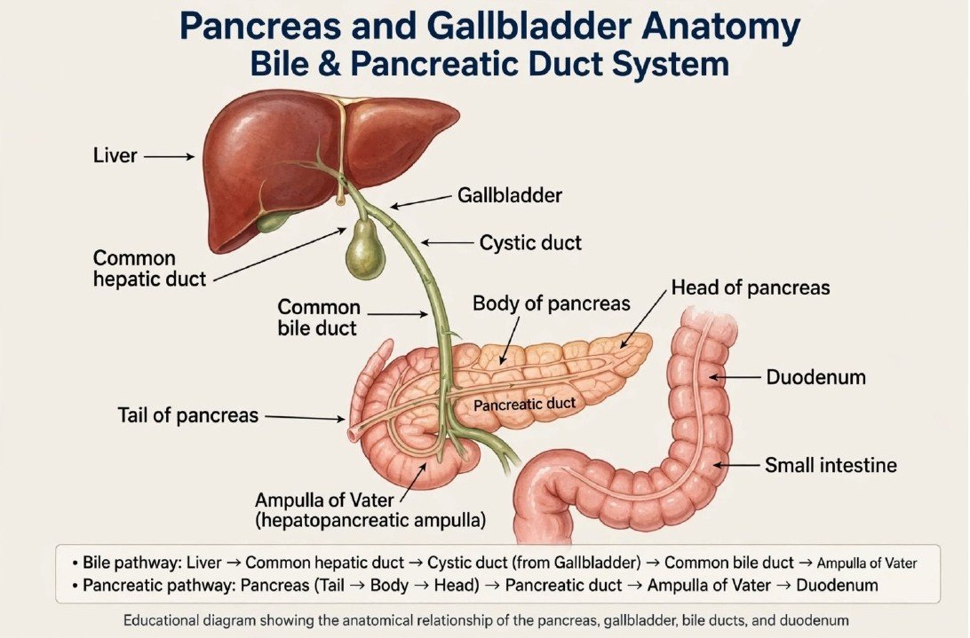
Pancreas, Gallbladder and Bile Ducts
This image is intended to explain anatomy, rather than a particular operation. The liver produces bile, which helps digest fats. The gallbladder stores and concentrates bile. The cystic duct joins the common hepatic duct to form the common bile duct, which carries bile towards the duodenum.
The pancreas supplies digestive enzymes through its duct and produces hormones, including insulin. Its head lies within the curve of the duodenum, while its tail extends towards the spleen. Understanding these structures helps explain treatments such as gallbladder removal and ERCP.
Illustration clarification: The pancreatic head and tail labels are reversed. The gallbladder is duplicated, several ducts are mislabelled, and bowel anatomy is inaccurate. Bile does not have to pass through the cystic duct before reaching thecommon bile duct.
Sources: NIDDK
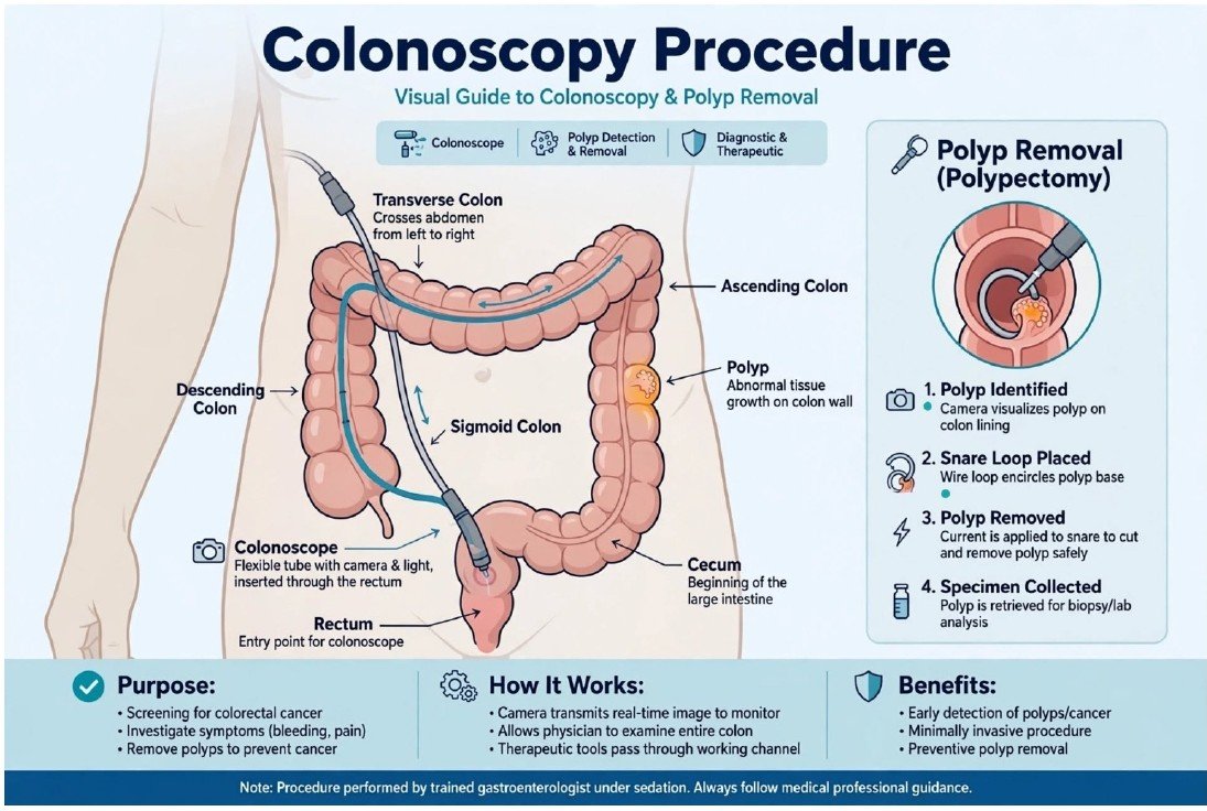
Colonoscopy and Polyp Removal
Colonoscopy uses a flexible camera passed through the anus and rectum to examine the lining of the large intestine. It is used for colorectal cancer screening and to investigate symptoms such as bleeding or persistent changes in bowel habits.
The clinician can take biopsies and remove suitable polyps during the examination. Polyp removal, called polypectomy, may use a wire snare with or without electrical current. Removed tissue is examined in a laboratory. Bowel preparation is needed, and sedation options depend on the patient and service.
Illustration clarification: Several colon segments are incorrectly labelled. The scope stays inside the bowel lumen after entering through the anus; the path drawn outside the colon is inaccurate. Electrical current is not required for every polyp removal.
Sources: NIDDK
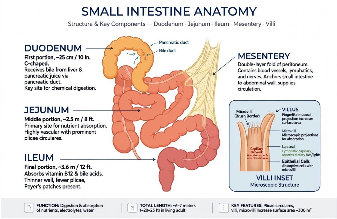
Small Intestine: Structure and Function
This is an anatomy illustration, not a surgical procedure. The small intestine has three sections: the duodenum, jejunum and ileum. It mixes food with digestive juices and absorbs most nutrients, along with water and electrolytes.
The duodenum receives bile and pancreatic secretions. The jejunum absorbs many nutrients, and the terminal ileum is important for vitamin B12 and bile acid absorption. Villi and microvilli increase the absorptive surface. The mesentery supports much of the intestine and carries vessels, lymphatics and nerves.
Illustration clarification: Some structures labelled as small intestine are drawn like colon, and the ileum arrow points towards large bowel. The stated lengths and surface area are not fixed values for every person.
Sources: NIDDK | Cleveland Clinic
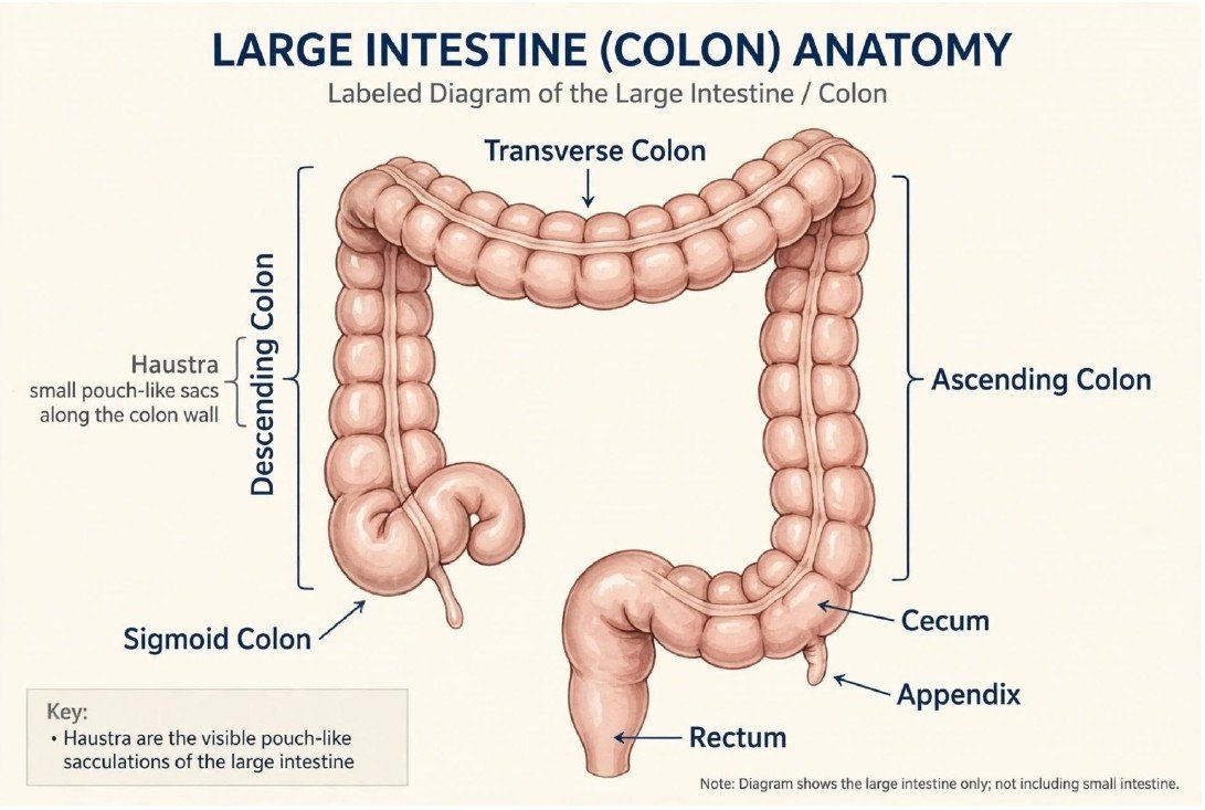
Large Intestine and Colon Anatomy
The large intestine absorbs remaining water and electrolytes, supports gut bacteria and forms and stores stool. In normal sequence, material passes from the ileum into the caecum, then through the ascending, transverse, descending and sigmoid colon, before reaching the rectum and anus.
The ascending colon is normally on the patient’s right and the descending colon on the left. The appendix is attached to the caecum. This image is intended as an anatomy overview; no operation is shown.
Illustration clarification: The ascending and descending colon labels are swapped relative to the depicted caecum and appendix. The sigmoid and caecum arrows are misplaced, and the drawing appears to include two appendix-like structures.
Sources: Cleveland Clinic
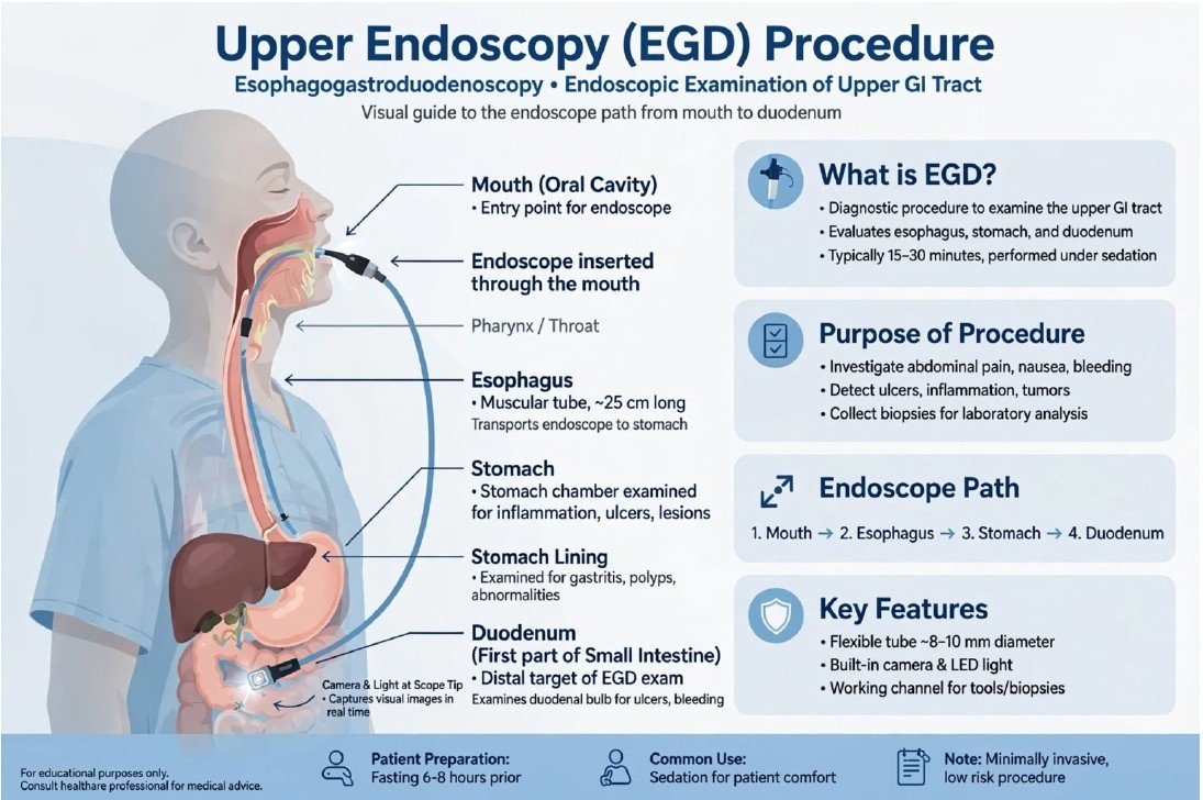
Upper Gastrointestinal Endoscopy (EGD)
Upper endoscopy uses a flexible camera passed through the mouth into the oesophagus, stomach and duodenum. It can investigate swallowing difficulties, upper abdominal symptoms, bleeding, ulcers and inflammation.
The clinician may take tissue samples or perform treatments such as controlling bleeding or widening selected narrow areas. It usually requires no external incision. Fasting and sedation instructions should come from the treating team, as requirements depend on the procedure and patient.
Illustration clarification: The endoscope must remain within the digestive tract. The drawing incorrectly suggests that part of the scope crosses outside the stomach and bowel. Procedure duration and fasting times are not identical for everyone.
Sources: NIDDK
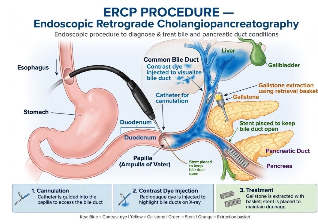
ERCP: Treatment of Bile and Pancreatic Ducts
Endoscopic retrograde cholangiopancreatography (ERCP) combines endoscopy and X-ray imaging, mainly to treat problems in the bile or pancreatic ducts. An endoscope travels through the mouth and stomach into the duodenum. A small catheter accesses the duct opening, allowing contrast imaging and treatment.
Depending on the problem, the clinician may remove bile duct stones, widen a narrowing, take samples or insert a stent to improve drainage. ERCP can cause pancreatitis, bleeding, infection or perforation, so it is used when the expected benefit justifies the risks. Stents may need planned removal or replacement.
Illustration clarification: The duct connections, scope path and duplicated pancreatic tissue are inaccurate. The endoscope stays within the digestive tract; instruments pass into the ducts through their opening in the duodenum. MRCP or endoscopic ultrasound may be preferred for diagnosis alone.
Sources: NIDDK
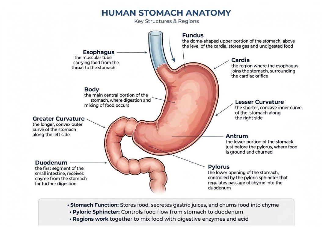
Stomach Anatomy and Digestion
The stomach receives food from the oesophagus, stores it temporarily and mixes it with acid and digestive enzymes. This produces a semi-liquid mixture called chyme, which passes through the pylorus into the duodenum.
Its main regions are the cardia, fundus, body and pyloric region, including the antrum. The lesser curvature is the shorter concave border; the greater curvature is the longer convex border. This image describes anatomy and does not show a specific surgery.
Illustration clarification: The cardia, fundus, pylorus and curvature arrows are misplaced. The cardia is at the oesophageal entry, the fundus forms the upper dome, and the pylorus is the outlet into the duodenum.
Sources: Cleveland Clinic